
FFIC1020
1
UNITED STATES MARINE CORPS
FORCE FITNESS READINESS CENTER
THE BASIC SCHOOL
24191 GILBERT ROAD
QUANTICO, VIRGINIA 22134
STUDENT OUTLINE
ANATOMY AND PHYSIOLOGY I
FFIC1020
FORCE FITNESS INSTRUCTOR COURSE
M02MN1T
APPROVED BY: LtCol (Ret) Shusko, J. C. DATE: 20190401 INT:______

FFIC1020
2
LEARNING OBJECTIVES
a. TERMINAL LEARNING OBJECTIVES
(1) Given a unit to train, supervise injury prevention to
increase Marine and unit readiness. (0919-TRNG-2004)
b. ENABLING LEARNING OBJECTIVES
(1) Without the aid of reference, identify the positions
of the human body without error. (0919-TRNG-2004d)
(2) Without the aid of reference, identify the planes of
the human body without error. (0919-TRNG-2004e)
(3) Without the aid of reference, identify the directions
of the human without error. (0919-TRNG-2004f)
(4) Without the aid of reference, identify the movements
of the human body without error. (0919-TRNG-2004g)
(5) Without the aid of reference, match the system of the
body to its function without error. (0919-TRNG-2004h)
(6) Without the aid of reference, identify major bones of
the human body without error. (0919-TRNG-2004i)
(7) Without the aid of reference, define the function of
ligaments within the human body without error.(0919-TRNG-200j)
(8) Without the aid of reference, identify major muscles
of the human body without error. (0919-TRNG-2004l)
(9) Without the aid of reference, define the function of
tendons within the human body without error. (0919-TRNG-2004m)
(10) Without the aid of reference, describe in writing,
human muscle contraction without error without error. (0919-
TRNG-2004n)
(11) Without the aid of references, define the different
muscle fiber types of the human body without error. (0919-TRNG-
2004p)
INTRODUCTION: There are three planes of movement for the human
body. The body is comprised of many various systems to support
these movements. In this class, you will learn the different

FFIC1020
3
ways the body moves and gain a basic knowledge of bodily
systems. This information serves as the foundational
information for other classes such as biomechanics, nutrition,
and practical application of exercise.
1. REFERENCE POSITIONS AND DIRECTION. When performing a
movement or exercise it is critical for the Force Fitness
Instructor (FFI) to understand the positions and directions of
the human body. The FFI will be better prepared to give
specific descriptions and education in exercise.
a. Positions of the Body
(1) Anatomical position. This is the standard position
from where all references and directions start: standing
upright, legs together and knees straight, toes pointing
straight forward, arms by the side, palms facing forward.
(2) Supine. The position of lying down, face up.
(3) Prone. The position of lying down, face down.
(4) Quadruped. The position with hand, knees, and feet
on the floor. Hands are under shoulders, knees are under hips,
and spine is neutral.
b. Planes and Sections
(1) Sagittal plane. The sagittal plane divides the body
into left and right. It passes through the body down the
middle. Movements in this plane are the up and down movements
like flexion and extension.
(2) Frontal plane. The frontal plane divides the body
into front and back. It passes through the side of the body.
Movements in this plane are lateral movements like abduction and
adduction.
(3) Transverse. This plane divides the body into top
and bottom. Movements in this plane are rotational like
internal and external rotation, or pronation and supination.

FFIC1020
4
c. Directional Terms
(1) Superior. Definition: above, toward the head or
upper part of the structure.
(2) Inferior. Definition: below, away from the head or
lower part of the structure.
(3) Anterior. Definition: in front of, front. The
sternum (breastbone) is anterior to the heart.
(4) Posterior. Definition: after, behind, nearer to or
back of the body.
Frontal (Or Coronal
Plane)
Sagittal (Median)
Plane
Posterior
Inferior
Transverse (Or
horizontal) Plane
Superior
Anterior

FFIC1020
5
(5) Medial. Definition: toward the mid-line, middle,
away from the side.
(6) Lateral. Definition: toward the side, away from the
mid-line.
(7) Proximal. Definition: near, closer to the origin.
(8) Distal. Definition: away from, farther from the
origin.
(9) Superficial. Definition: situated near the surface
of the body.
(10) Deep. Definition: describes structures that are
away from the surface of the body. The ribs are deep to the
skin.
d. Movements
(1) Flexion. A movement which decreases the angle at
the moving joint. This movement occurs in the sagittal plane.
(2) Extension. A movement which increases the angle at
the moving joint. The opposite movement of flexion. This
movement occurs in the sagittal plane.
(3) Abduction. Taking the limb away from the central
line of the body. This movement occurs in the frontal plane.

FFIC1020
6
(4) Adduction. Taking the limb towards the central line
of the body. This movement occurs in the frontal plane.
(5) Rotation. This movement includes any twisting
motion. This movement occurs in the transverse plane. Joints
which permit rotation include the shoulder and hip as an
example.
(6) Circumduction. A combination of all movements
above. It is possible at the ball and socket joint, as if you
were to draw a circle with your arm.
(7) Pronation. Pronation is the movement of turning the
palm over to face downwards from the anatomical position.
(8) Supination. Supination is the opposite movement of
pronation, of turning the palm up or forwards into an anatomical
position.

FFIC1020
7
2. SYSTEMS OF THE BODY. The human body is divided into several
systems and each has specific functions. This will become base
knowledge of exercise physiology in future classes.
a. Endocrine System. The endocrine system regulates body
activities by releasing hormones, which are chemical messengers
transported in blood from gland to specific organ.

FFIC1020
8
(1) Pituitary Gland. During exercise, the pituitary
gland releases human growth hormone, which tells the body to
increase bone, muscle, and tissue production.
(2) Thyroid Gland. When you start exercising, the
thyroid gland (located at the base of the neck) sends out
hormones that regulate the body’s temperature, heart rate and
blood pressure. It also regulates the alertness and focus
needed to work at a high intensity.
(3) Adrenal Gland. Located at the top of the kidneys,
the adrenal glands are responsible for the release of cortisol
into the bloodstream. Cortisol levels control blood pressure,
glucose and act as an anti-inflammatory agent. The adrenal
glands also releases aldosterone, a hormone that regulates
hydration levels, the speed of the heart and the strength of
contractions. It also turns stored carbohydrates into energy.
(4) Pancreas. Insulin regulates glucose, or blood
sugar, by transporting it to the muscles and tissue that use
glucose for energy. Excessive insulin in the blood reduces your
sensitivity to insulin and can cause diabetes, which is also

FFIC1020
9
linked to overweight and obesity. Exercise improves insulin
sensitivity and reduces the reliance on insulin injections.
b. Integumentary System. The integumentary system protects
the body, detects sensation, and produces vitamin D. Most often
we relate this system to sweating (evaporation), skin injuries
(blisters) and infections (ringworm, dermatitis) during
training.

FFIC1020
10
c. Lymphatic System. The lymphatic system returns proteins
and fluid to blood and carries lipids from intestines to blood.
The lymphatic system is also involved with fighting bacteria and
infection. It is a “return” system of fluid.

FFIC1020
11

FFIC1020
12
d. Digestive System. The digestive system achieves
physical and chemical breakdown of food, absorbs nutrients, and
eliminates waste. Most exercise has a positive effect on the
digestive system helping to quell appetite and increase
metabolism. Some endurance events sometimes cause competitors
to have an upset stomach and diarrhea.
(1) Stomach. The stomach acts as a reservoir for the
food where it may remain between 2 and 6 hours. Here, the food
is churned over and mixed with various enzymes, hydrochloric
acid, and other chemicals; all of which are secreted further
down the digestive tract. The stomach has an average capacity
of 1 liter and is capable of considerable distension. When
expanding, stimuli is sent to the hypothalamus which is the part
of the brain and nervous system which controls hunger. The wall
of the stomach is impermeable to most substances, although does
absorb some water, electrolytes, certain drugs, and alcohol.
(2) Small intestine. The small intestine measures about
7 meters. Both the bile (from the liver) and pancreatic (from
pancreas) ducts open into the small intestine together. The
small intestine provides a vast surface area where further
absorption takes place. There is a large blood supply to this
area, ready to transport nutrients to the rest of the body.
(3) Pancreas. The pancreas has two main functions: to
produce enzymes to aid the process of digestion and to release

FFIC1020
13
insulin directly into the blood stream for the purpose of
controlling blood sugar levels.
(4) Liver. The liver, which acts as a large reservoir
and filter for blood, has several important functions: secretion
of bile to the gall bladder (break down fats), metabolism of
carbohydrates, protein and fat, storage of glycogen ready for
conversion into glucose when energy is required, and the storage
of vitamins.
e. Urinary System. The urinary system has multiple
functions, such as excreting toxins and metabolic by-products,
maintaining the body's fluid and acid-base balance; regulating
electrolyte levels, and secreting several important hormones.

FFIC1020
14
f. Reproductive System. The reproductive system releases
hormones and creates human life.
3. SKELETAL SYSTEM IN DEPTH.
a. Functions. The skeletal system supports and protects
vital organs, provides attachment, stores minerals, and gives
rise to blood cells.
(1) Protects vital organs. For example, the rib cage
will protect the vital organs of the heart and lungs. Your
cranium protects the brain.
(2) Provides attachment and structure. Muscles attach
to the bone to create movement. Bones give the human body shape
and the base frame work.
(3) Stores minerals. Bones store key minerals such as
calcium and magnesium.
(4) Ligaments. While “ligaments” are considered a type
of tissue and not classified in the skeletal system, it is worth
noting that bones are jointed together using ligaments. A
common knee ligament is the ACL or Anterior Cruciate Ligament.
More will follow in the Common Injuries lecture.

FFIC1020
15
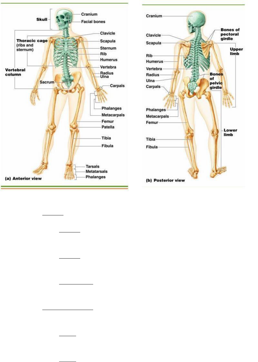
b. Anatomy. The major bones of the body are labeled in the
second diagram below: skull, cranium, jaw bone, ribs, scapula,
humerus, radius, ulna, carpals, metacarpals, phalanges,
vertebrae, sacrum, pelvis, femur, patella, tibia, fibula,
tarsals, metatarsals, phalanges.
FFIC1020
16
(1) Axial. The axial skeleton has three main parts.
(a) Skull. The skull holds the brain in a liquid
suspension.
(b) Spine. The spine is composed of individual
bones separated by disc cartilage, and protects the spinal cord.
(c) Rib cage. The ribs protect vital organs with 12
pairs of ribs.
(2) Appendicular. The appendicular skeleton includes
the body’s limbs and girdles.
(a) Arms. The arms are used for lifting and
carrying.
(b) Legs. The legs are used for movement and
propulsion.

FFIC1020
17
4. MYOFASCIAL SYSTEM IN DEPTH. The myofascial system includes
all the muscles of the body and the fascia (encasement tissue)
that surrounds it. It will be necessary to identify the major
muscles of the body.
a. Functions. There are several main functions of the
myofascial system.
(1) Body movements. This system coordinates total body
movements such as running and walking. Also, localized motions
such as writing or nodding the head.
(2) Stabilizing body positions. The myofascial system
increases the stability of joints and maintains body positions
such as sitting. The myofascial system help keep joints within
their intended space and stabilize the joint during motion.
(3) Moving substances in the body. Cardiac muscle moves
blood through the body. Smooth muscles move food and substances
in the gastrointestinal tract. Skeletal muscle contractions
promote blood flow.
(4) Providing heat. As muscle contracts, it produces
heat. As temperature increases, muscles help release the heat.
Involuntary muscle contractions such as shivering can increase
rate of heat production.
(5) Tendons. It is worth noting that muscles attach to
bones using tendons. A common knee tendon is the patella
tendon. It is the same tendon doctors use to test the knee
reflex. More will follow in the Common Injuries lecture.

FFIC1020
18
b. Anatomy. There are numerous muscles in the human body,
the picture below serves as a reference of the individual
muscles. For the purpose of this class we will group the major
muscles together.
(1) Major muscles of the lower body: The major muscles
of the lower body are: hamstrings, quadriceps, gluteus maximus,
gastrocnemius, and soleus muscles.

FFIC1020
19

FFIC1020
20
(2) Major muscles of the trunk: The major muscles of
the trunk body are: external oblique, internal oblique,
transverse abdominis, and rectus abdominis. You can see the
pectoralis major at the top of the picture. The deep muscles of
the back (posterior view) can be grouped as the erector spinae
muscle group. The more superficial muscles of the back are
latissimus dorsi and trapezius. The deltoid muscle is better
viewed from the lateral view but the posterior muscle fibers can
be seen here.

FFIC1020
21

FFIC1020
22
(3) Major muscles of the upper body. The main muscle of
the upper arm are the biceps (anterior) and triceps (posterior).
The main muscles of the lower are going to be generalized in the
forearm. The distal anterior arm muscles are the “wrist
flexors,” and the distal posterior arm muscles are the “wrist
extensors.”
c. Connective Tissue. Connective tissue (fascia) surrounds
and protects muscle tissue.
(1) Fascia. Fascia is defined as a sheet or broadband
of fibrous tissue deep to the skin, and surrounds muscles and
other types of organs in the body.
(2) Fascia in Muscle. Fascia separates muscle from skin
and provides a pathway for nerves and blood vessels to enter and
exit the muscles. It allows free movement of muscles, carries
nerves, blood and lymphatic vessels, and fills the space between
muscles. Adhesions may occur naturally with use, disuse,

FFIC1020
23
exercise, or injury. Fascia can restrict overall mobility and
be a part of force production in muscle strength.
d. Muscle contractions
(1) Motor unit. A motor unit is the nerve fiber and all
the muscle fibers it innervates. A single motor unit has an
average of 150 muscle fibers. Force production = recruitment of
more motor units.

FFIC1020
24
(2) Sarcomere. A sarcomere is the smallest functional
unit of a muscle fiber. Characterized from Z-line to Z-line
(below). The sarcomere is made up of thick and thin filaments.
Thick and thin filaments form cross bridges to shorten the
muscle fiber.
(3) Contraction step 1 (release & energize). The first
step, in the cycle involves the binding of a molecule of
adenosine triphosphate (ATP) to the myosin head. When the ATP
binds, the myosin head releases from the thin filament and is
ready to undergo the contraction cycle.
(4) Contraction step 2 (slide forward). In the second
step, the myosin head breaks down the bound ATP molecule into
ADP and P, but instead of releasing the products, the myosin
head holds onto the ADP and P. This causes the myosin head to
make its first change in shape. It enters the "cocked" position
as it slides forward.
(5) Contraction step 3 (bind & excited). The third step
is when the excited myosin head binds to the actin strand (thin
filament). When this occurs, the P that was bound to the myosin
head is released. When it is released, the actin binding site
on the myosin molecule is exposed. The actin and myosin head
are tightly held together for the remainder of the cycle. While

FFIC1020
25
the release of the P molecule causes a change in shape of the
myosin head, it is still in the excited cocked state in this
step.
(6) Contraction step 4 (power stroke). The final step is
when the myosin head releases the bound ADP. When this happens,
the myosin head leaves its excited state and returns to the
position where it began. But this time, the myosin head is
tightly bound to the thin filament, so when the myosin head
shifts, it pulls the thin filament with it. This shift of the
actin and myosin is called the "power stroke". In this step,
the energy that was generated by breaking down ATP, and stored
in the excited "cocked" position of the myosin head is finally
used to contract the muscle. After the "power stroke", the
cycle is completed when the myosin head binds to a new ATP
molecule, if ATP is available, causing the myosin head to let go
of the actin strand.

FFIC1020
26
e. Muscle fiber types
(1) Slow Twitch. Slow twitch fibers develop force
slowly and relax slowly.
(a) Type I. Type I fibers (slow twitch) are
described as the following: slow, withstands fatigue, efficient,
aerobic capacity. Limited in maximum force development and has
low anaerobic power. Example: a professional marathon runner
has more Type I fibers.
(2) Fast twitch. Fast twitch fibers develop force
quickly and relax quickly.
(a) Type IIa. Type IIa fibers are described as:
fast, inefficient and fatigable with moderate aerobic power.
They create rapid force development and have high anaerobic
power. You can summarize this fiber type as an “in between”
muscle fiber not truly fast or slow. Type IIa fibers have a
higher capacity for aerobic power compared to IIx due to an
increase of capillaries surrounding the muscle. Example: Running
the (800M) will use the type IIa fibers.
(b) Type IIx. Type IIx fibers are described as:
fast, inefficient and fatigable with low aerobic power. They
create rapid force development and have high anaerobic power.
These are considered “true” fast twitch fibers.

FFIC1020
27
SUMMARY: While reviewing the systems individually, you can see
how each system is integrated with other systems. You may now
be able to see how nutrition and supplements can affect the
human body’s system. Finally, the basic understanding of
anatomy and physiology will be the basis of kinesiology and
biomechanics.
REFERENCES:
Gray's Anatomy, 13th Ed. Clemente, 1985. GRAY'S ANATOMY
Marine Corps Physical Fitness Program MCO 6100.14_
Jack H.; D. L. Costill.Physiology of Sport and Exercise, 2nd
Edition, Wilmore,ISBN:0736062262
Risk Management (RM) MCO 3500.27_
